PROJECTS
Surgery through advanced imaging
From scan to strategy: how imaging drives personalised surgery
Advanced imaging helps us plan surgeries that are more accurate, safer, and built around the individual. We use medical imaging to support more accurate diagnosis, surgical planning, and evaluation of outcome. By creating detailed 3D models of bones and joints, we help tailor surgical strategies to each individual patient.
How we use imaging for better patient outcomes
Modern hip and knee surgeries use 3D-imaging and planning tools to choose the best implant size, position, and fit for each patient. Our lab supports this approach through:We specialise in three main areas:
POST-SURGICAL EVALUATION
Comparing pre-operative plans to post-op outcomes
Using imaging to improve future planning and precision
TRACKING IMPLANT PERFORMANCE OVER TIME
Monitoring how implants behave inside the body
Detecting early signs of movement, wear, or failure beyond simple revision rates
PATIENT-SPECIFIC SURGICAL PLANNING
Using CT/MRI to create 3D bone models
Planning implant size and position before surgery
Producing Patient Specific Instrumentation (PSI) to guide the surgeon in real time
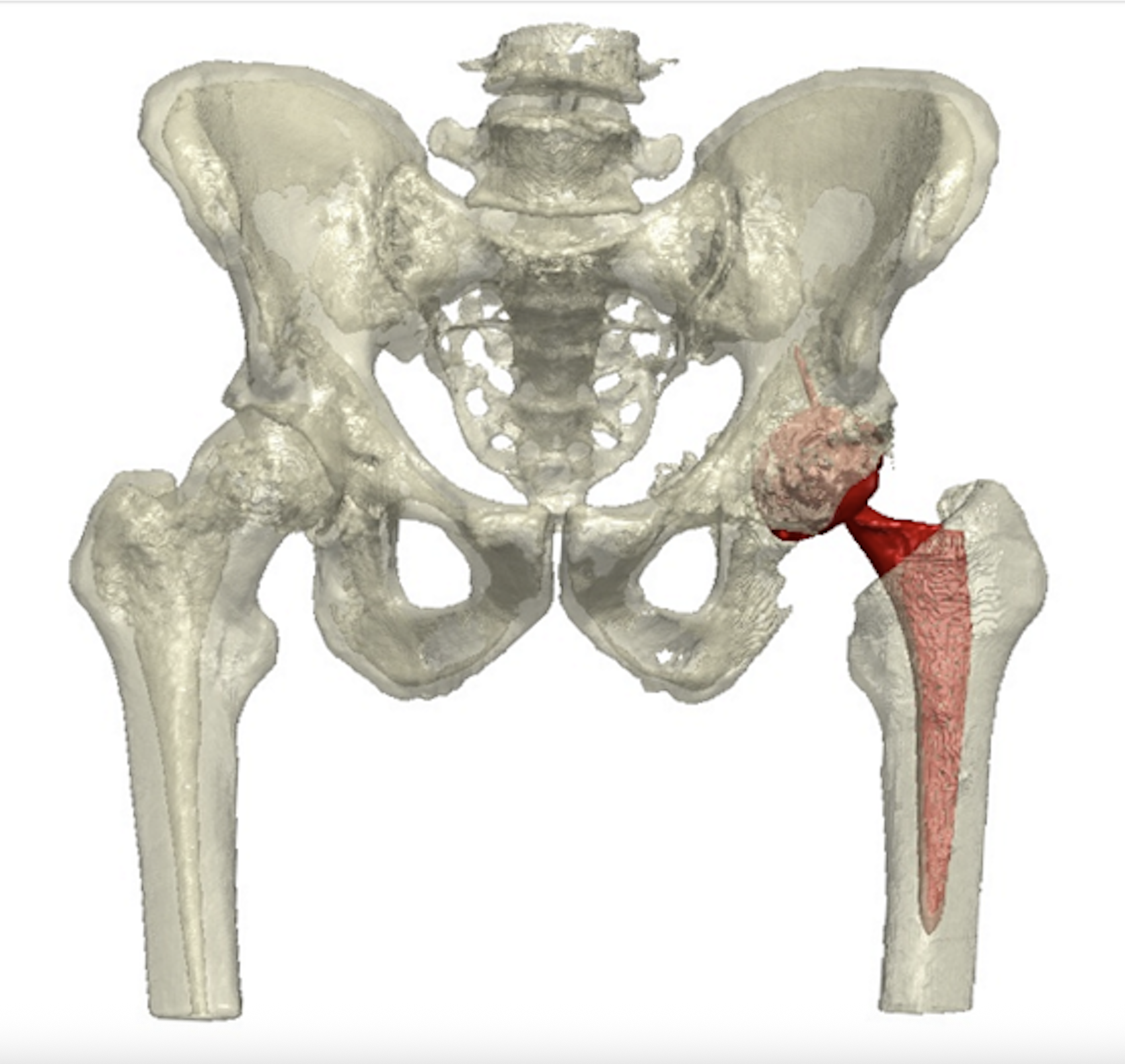
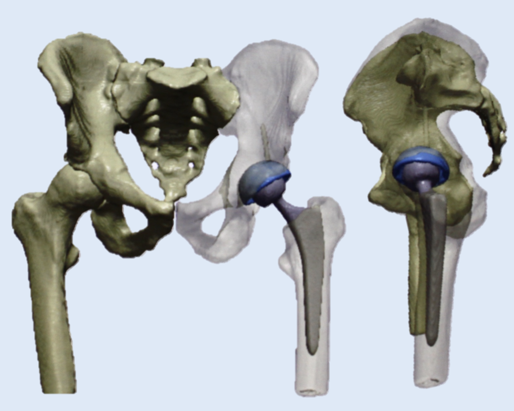
3D pelvis and hip implant demonstrating the acetabular and femoral component position and orientation and illustrating the restoration of centre of rotation and leg lengths.