CASE STUDY
Understanding how the pelvis moves in 3D
Every patient’s pelvis moves differently, and this variation affects the ideal orientation of a hip implant. Understanding an individual’s spinopelvic motion helps surgeons position implants more precisely and tailor the surgical plan to the patient.
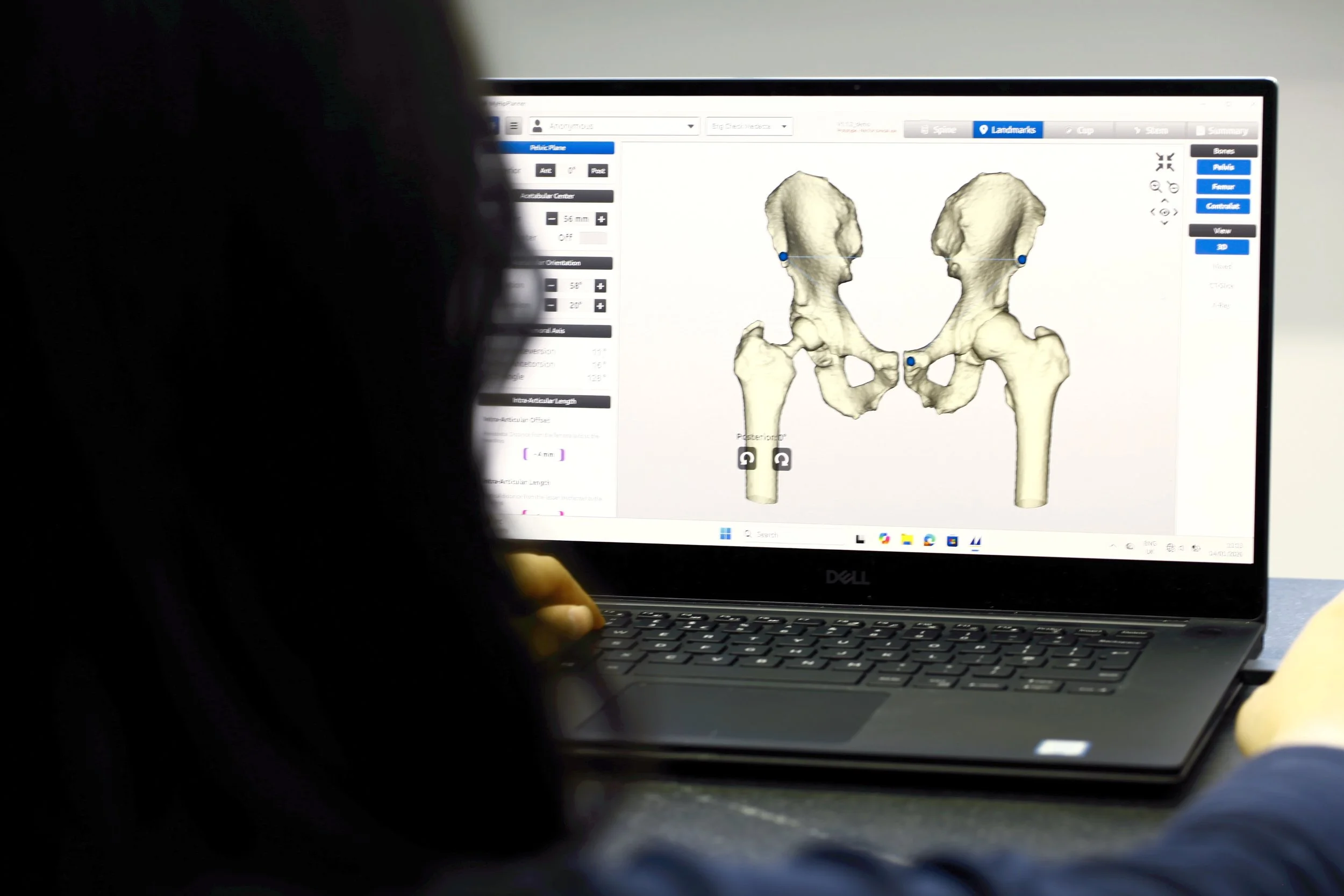
Traditional CT scans provide excellent anatomical detail, but they are taken with the patient lying down and cannot show how pelvic tilt changes when standing, sitting, or moving. This makes it difficult to understand each patient’s functional posture — a factor that can significantly influence implant alignment and stability.
The challenge
Most CT scans are taken with the patient lying down (supine). While CT gives excellent detail of the bones, it doesn’t show how the pelvis behaves in real-life positions—like standing or sitting—which are critical for planning hip surgery.
Our solution: a hybrid approach
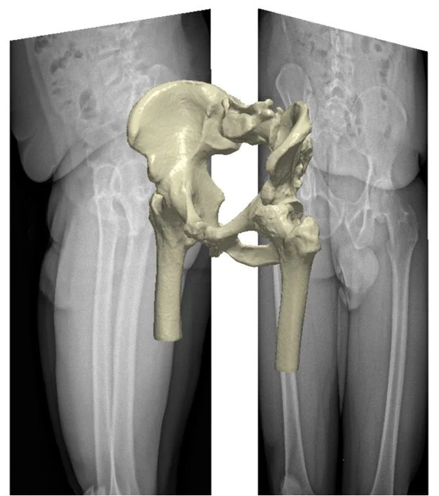
We combine detailed 3D models from CT scans with upright EOS imaging to capture the pelvis in real-life, weight-bearing positions. By aligning (registering) the CT-based model with EOS images, we can measure pelvic tilt and orientation in functional postures. This gives a more complete, 3D picture of how the pelvis behaves when the patient moves between positions that matter in daily life.
Why it matters
Everyone’s pelvis moves differently when changing posture. Understanding this movement helps surgeons decide the best position for the implant during hip replacement. This technique supports a more personalised approach to surgery, reducing the risk of complications and improving outcomes.