
CASE STUDY
Time to standardise CT protocols for 3D planning of hip and knee replacement
Computed tomography now plays a central role in three dimensional planning for joint replacement. It underpins implant sizing, version assessment, alignment analysis and the growing use of robotic and navigated workflows. While the value of CT based planning is widely accepted, far less attention has been paid to how these scans are acquired.
Unlike many other aspects of arthroplasty, CT protocols for preoperative planning remain highly variable. In daily practice, radiographers are often asked to follow vendor specific instructions that differ markedly between implant systems. These protocols frequently lack critical technical detail, leaving key decisions to be made at the scanner console. The consequence is wide variation in image quality and, more importantly, radiation dose delivered to patients.
This variability is unnecessary and increasingly difficult to justify.
Radiography is too often a source of unintended variability in an otherwise highly standardised surgical planning process. The data is clear: current manufacturer protocols vary widely and frequently omit key parameters, leaving radiographers to guess at dose and quality trade-offs. The orthopaedic community; surgeons, radiographers, physicists and vendors alike must now unite around a common set of harmonised CT standards that protect patients and advance practice
The challenge
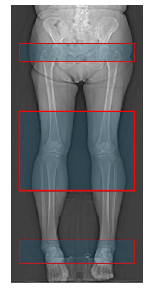
CT protocols vary widely between vendors and institutions, often lacking clear guidance on dose, noise or key technical parameters. This leaves radiographers to make critical decisions at the scanner, resulting in unnecessary variation in image quality and, more importantly, patient radiation exposure.
Our solution
The anatomical and imaging requirements for arthroplasty planning are consistent, regardless of implant brand. Recognising this, harmonised, vendor-neutral CT protocols can replace fragmented, incomplete guidance. Procedure-specific protocols that clearly define scan coverage and acquisition parameters remove ambiguity, support radiographers, and enable reliable low-dose imaging that is fit for purpose. Standardisation does not mean a single one-size-fits-all scan, but a coherent framework grounded in clinical need rather than commercial convention.
Why it matters
Standardised CT protocols improve safety, efficiency and reproducibility, while providing a stable foundation for advanced planning software and robotic systems. Harmonisation is not a constraint on innovation—it is a prerequisite for safer, more reliable arthroplasty care.