
CASE STUDY
Understanding variation inside the femur
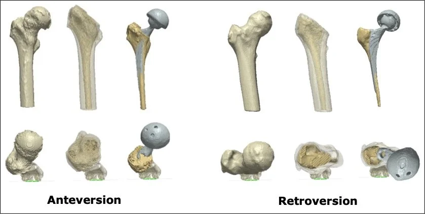
When surgeons plan an uncemented hip replacement, they rely on the shape of the inner femoral canal to achieve a secure, stable fit. However, most conventional planning focuses on the outer shape of the bone, assuming that the inside follows a predictable pattern. Our research shows that this isn’t always the case.
Even when the external anatomy looks similar across patients, the internal canal can vary greatly in size, curvature, and orientation. These differences can affect how well an implant grips the bone, how forces are distributed, and how long the implant lasts. Without understanding this internal variation, surgeons may unintentionally select or position an implant suboptimally.
The challenge
When placing an uncemented hip implant, the shape of the inner femoral canal is crucial for stability. But most planning is based on the bone’s outer shape.
Our solution: shape modelling
We used statistical shape modelling (SSM) to study how the inner canal of the thigh bone varies across individuals. We found substantial variation—even among people with similar external anatomy. SSM allowes us to:
• Quantify natural differences in canal size and shape
• Identify subtle patterns that may influence implant stability
• Predict internal geometry more accurately based on external anatomy
Why it matters
A clearer understanding of internal femoral variation helps surgeons plan more personalised and precise hip reconstructions. It supports better implant selection, more accurate positioning, and improved long-term stability—especially in uncemented hip replacements where secure bone–implant contact is essential.