
CASE STUDY
Understanding Acetabular Variation in Total Hip Arthroplasty
Accurate acetabular cup positioning is critical to the success of total hip arthroplasty (THA), influencing joint stability, range of motion, wear, and long-term implant survival. Conventional surgical planning relies on population-based “safe zones” for cup inclination and anteversion. However, growing evidence shows that pelvic and acetabular anatomy varies substantially between individuals and between sexes.
The acetabulum is not a uniform structure: its orientation, depth, and spatial relationship to the pelvis differ widely across patients. These anatomical differences are often not fully accounted for in standard planning workflows, increasing the risk that a “correctly positioned” implant may still perform poorly for a given patient.
Failure to account for acetabular anatomical variation can lead to suboptimal implant positioning, even when conventional alignment targets are met. This mismatch between implant orientation and native anatomy is a recognised contributor to instability, impingement, abnormal wear, and early revision following total hip arthroplasty.
The challenge
The main challenge is quantifying and understanding acetabular anatomical variation in a clinically meaningful way.
Native acetabular orientation varies significantly across individuals
Sex-specific differences further increase variability
Traditional safe zones do not account for these variations
Surgeons lack objective tools to relate individual anatomy to optimal cup positioning
Our solution
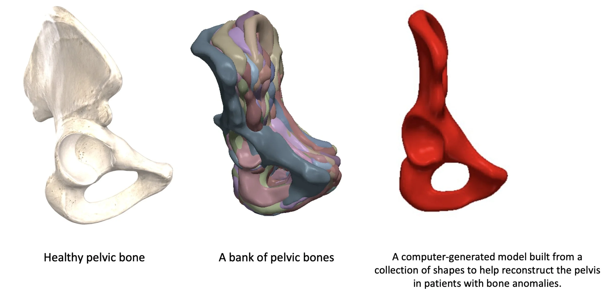
We developed a computational, data-driven approach to characterise acetabular variation using three-dimensional imaging and statistical shape modelling.
CT-derived 3D pelvic models were used to capture true anatomical geometry
Sex-specific statistical shape models quantified dominant modes of acetabular and pelvic variation
Principal component analysis identified how acetabular orientation contributes to overall shape variability
The models revealed systematic differences in acetabular inclination and version across individuals
This approach enables acetabular orientation to be interpreted in the context of the patient’s native pelvic morphology, rather than against a fixed population average. By integrating anatomical variation into surgical planning, this work supports a shift toward personalised acetabular cup positioning, moving beyond one-size-fits-all safe zones.
Why it matters
By objectively characterising how acetabular orientation varies between individuals and between sexes, this work highlights the limitations of population-based safe zones and reinforces the need for patient-specific planning. Incorporating anatomical variation into pre-operative decision-making has the potential to improve functional outcomes, reduce complications, and support more consistent surgical performance across diverse patient populations.